This month, we were fortunate enough to take a break from our normal day to day lab work and host a group of 25 high school students from the Glenbard East High School of Lombard, Illinois and the Hockaday School of Dallas, Texas. We reinforced Nipam’s colorful lectures on development with zebrafish timelapses, antibody stains, and butterfly wings.
The students were able to watch their zebrafish embryos develop and Jenny even showed a GFP Fli line, in which they got to see the heart beating and blood moving throughout the fish!
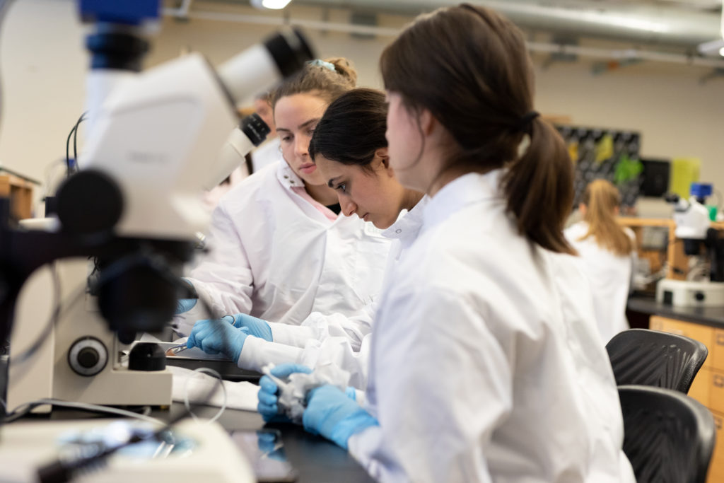
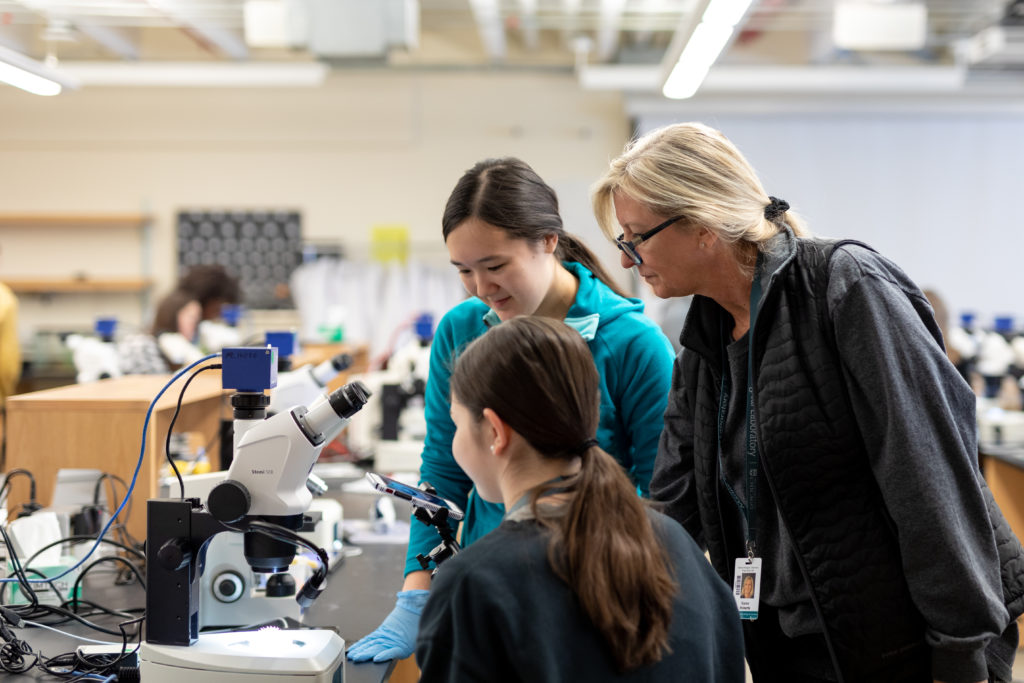
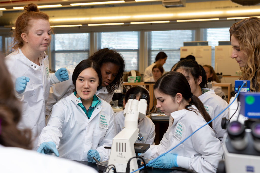
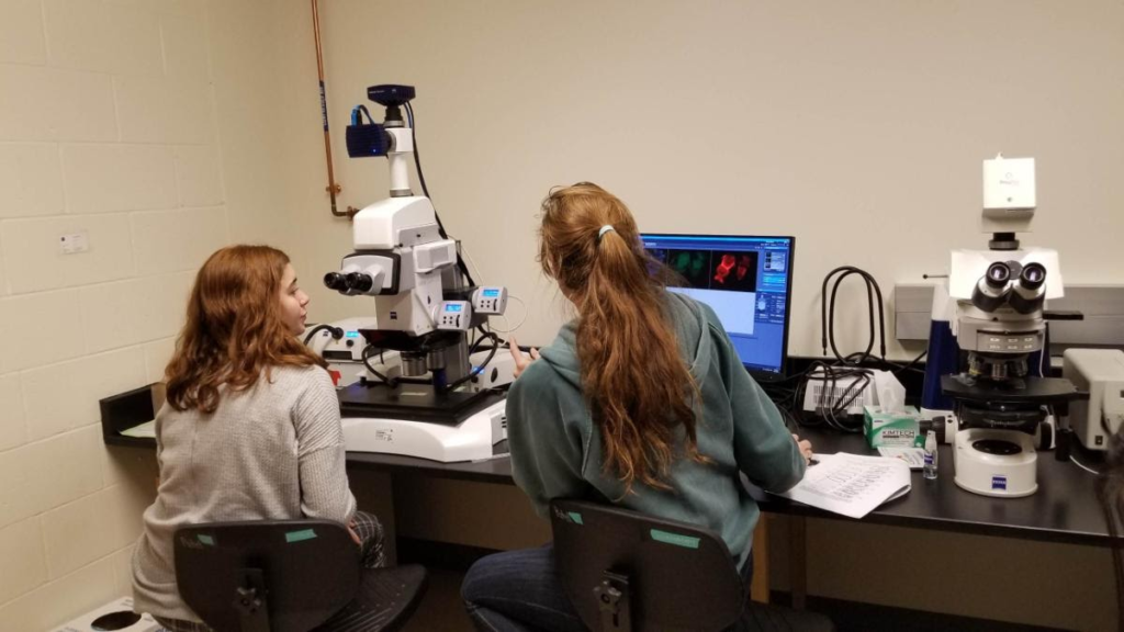
We also taught the students about antibody staining and had them complete their own stains on fly, zebrafish, squid, and cuttlefish embryos. They joined us in our lab space to learn how to mount their antibody stains under dissecting microscopes and under fluorescence on the Zeiss AxioZoom V16 and Zeiss Imager A.1 microscopes…
…they were very amused at our use of NYC Pink Promenade nail polish to seal down our cover slips. Besides the fact that the polish is an amazing, vibrant pink, we use this polish because it does not fluoresce under the lasers of the confocal and is the perfect consistency, as it does not spread out all over the slide. With that being said, we have been having a hard time finding this Pink Promenade for sale anywhere, so Erin has purchased a wide range of different NYC polishes that two of the students so graciously tested out for us, returning the next day with detailed notes on the consistencies, colors, and dry times (Thank you Paige and Helena!). Who said fashion isn’t important for science?!


Once the students had their stains mounted, they accompanied Nipam on the Zeiss LSM 880 to learn how confocal microscopy works and take their own images to process in ImageJ.


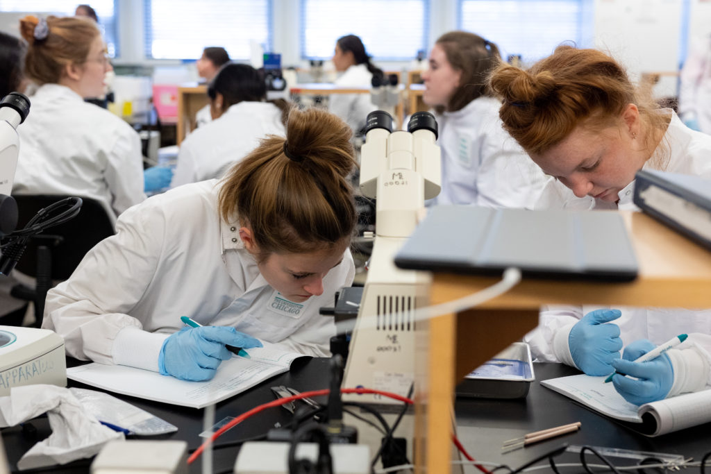
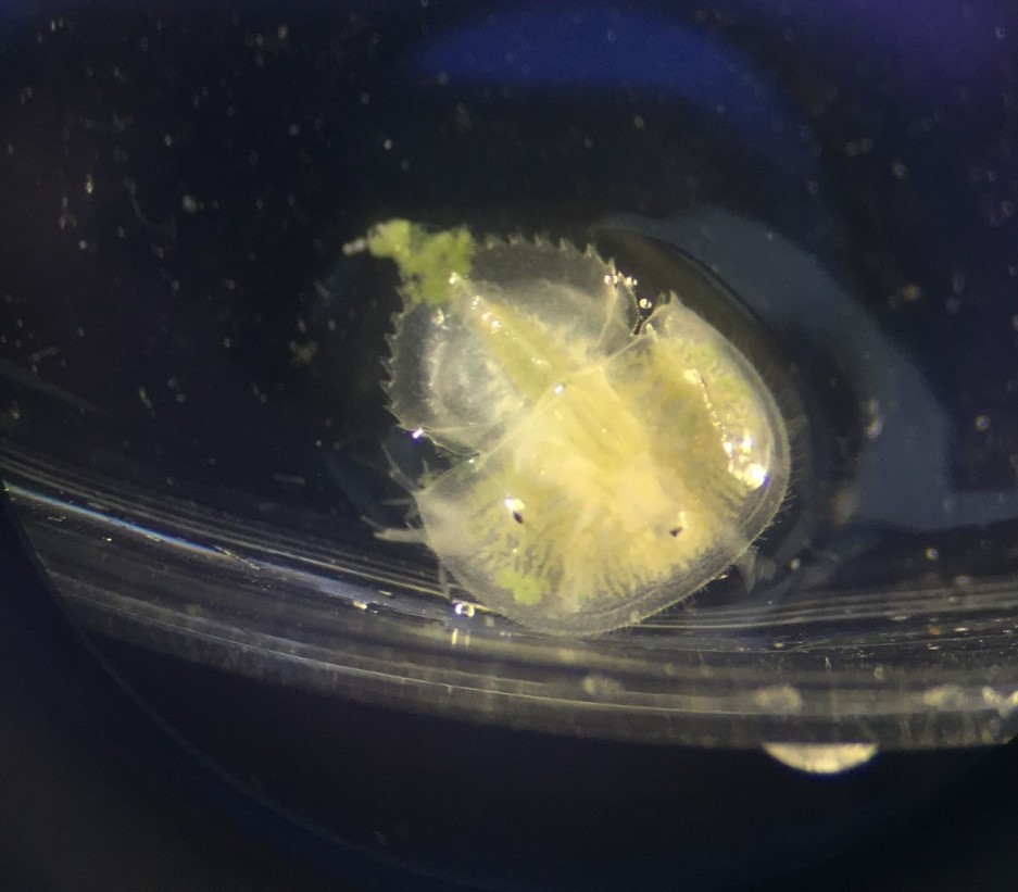
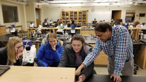
Top left: Students taking images of their antibody stains on the confocal with Nipam. Bottom left: Students analyzing and processing their images from the confocal on ImageJ. Images taken by Glenbard East chaperone Marisa Abrams. Right: Squid embryo confocal image taken by Tia Hsieh (Hockaday); Imaged on a Zeiss LSM 880 and processed using ImageJ. Squid embryos from the Mariculture team in the Marine Research Center at MBL.
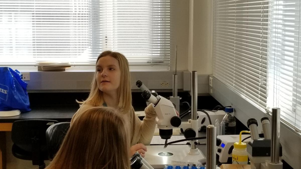
Last, but definitely not least!, the students were able to explore the beauty of butterfly wings. Through a small experiment with acetone and xylene, they examined different species of butterflies to understand if the scales exhibit structural colors, pigmentary colors, or both! They even got a more close-up view of the wing scales with Kyle on the Keyence microscope in our lab.


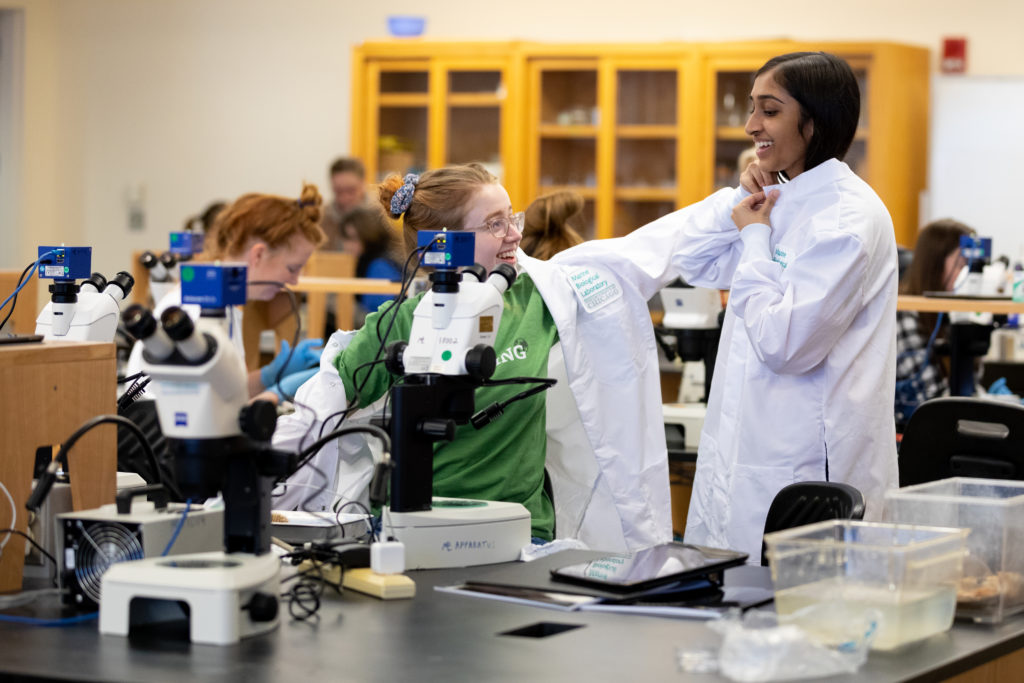
In the chaos of finishing up high school and applying to colleges (goodluck girls!), we were so pleased with the students’ enthusiasm throughout the week! It is always a pleasure working with young, bright-eyed students, as we can only hope that spending time at the Marine Biological Laboratory will help inspire future generations of scientists to come!
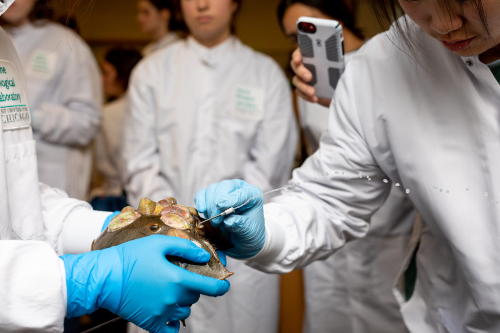
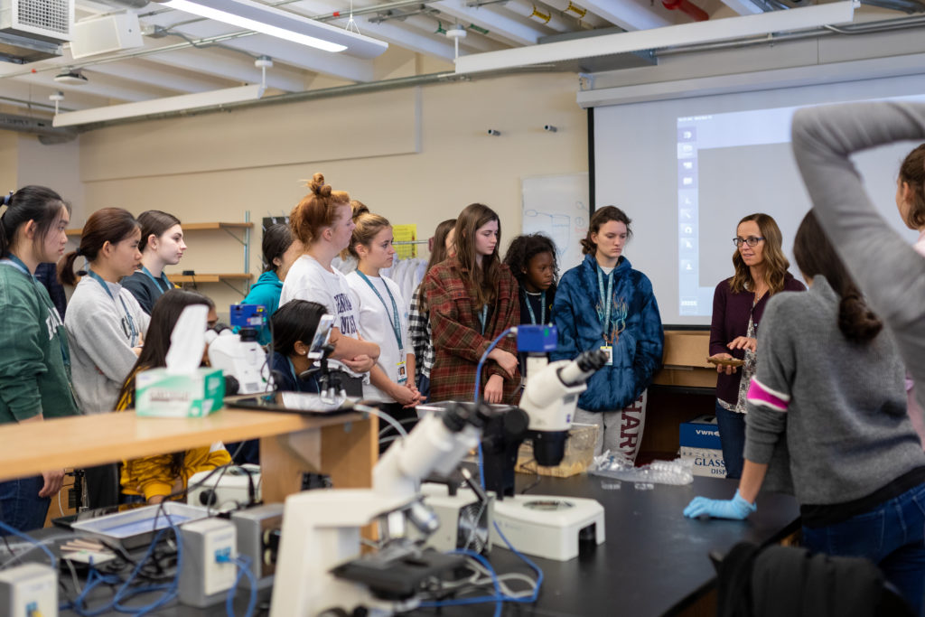
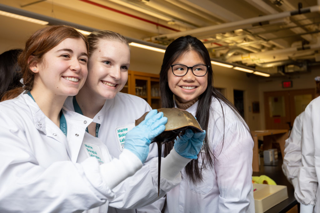
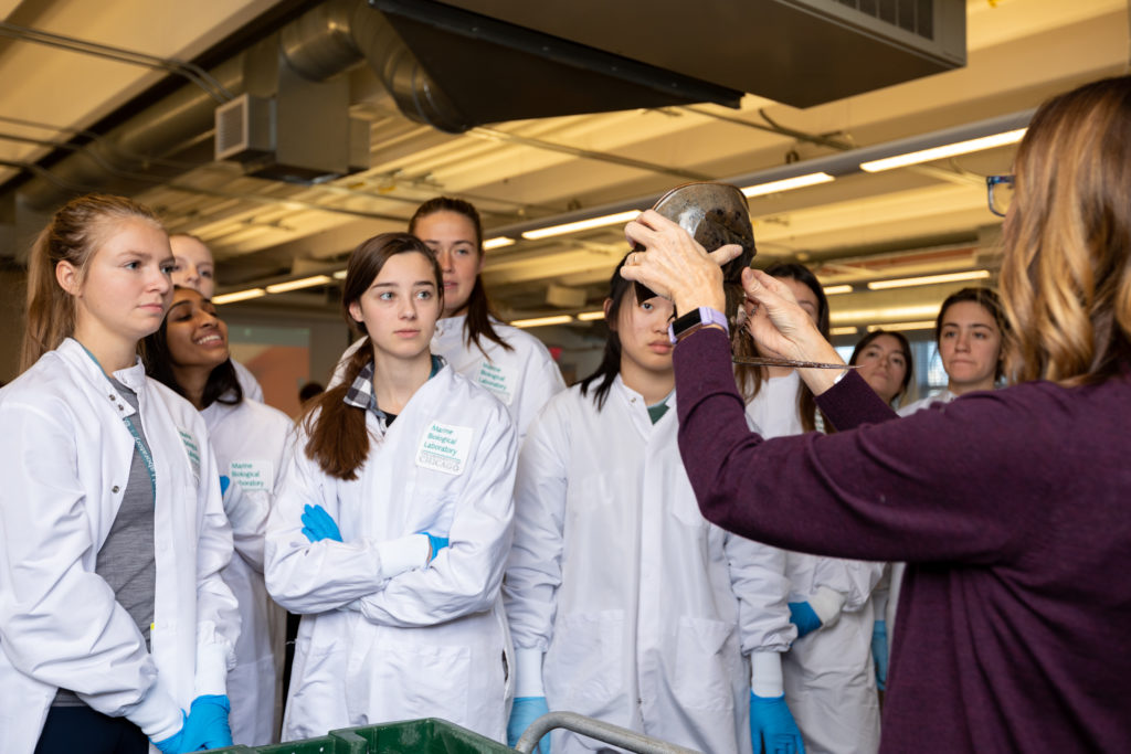
In addition to the development section of the course, the students also explored local marine life with our veterinarian, Lisa Abbo. The following pictures were taken by Dee Sullivan (https://www.minfinphotography.com/):